The heart is an awesome piece of engineering. It pumps our blood at an incredible rate. It can withstand a great deal of strain and pressure and still manage to do its job! So how do we know if it is doing its job well or not? We can’t physically examine our heart, but we can observe the effects it has.
ne way of looking at heart health is through an electrocardiogram (ECG). This course will take you through the electrophysiology of the heart, reading the electrical stimuli it produces and the changes that occur when it’s not working efficiently.
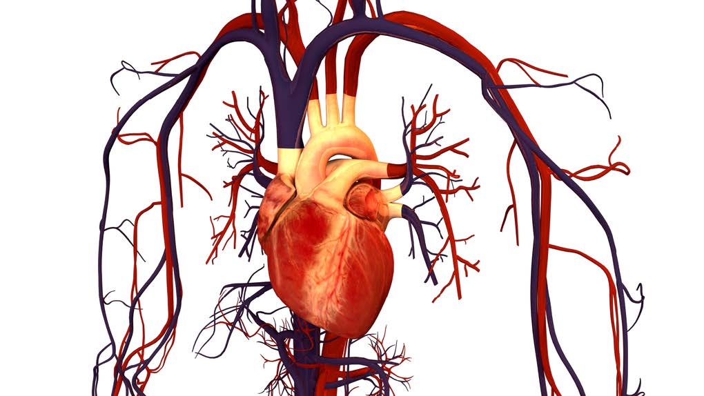
Before looking at the electrophysiology of the heart, let’s review the basic cardiac anatomy. The adult heart is approximately the size of your fist and sits in the left side of the thoracic cavity in front the lungs. The heart is orientated in the chest rotated at about 30 degrees to the left lateral side (Kaelber, 2013). The heart is a mechanical pump driven by electrical activity.
The heart has four chambers:
- Right atrium (RA)
- Right ventricle (RV)
- Left atrium (LA)
- Left ventricle (LV)
The two atria are the smaller chambers and the two ventricles are the larger chambers of the heart. The right ventricle is the most anterior structure of the heart. The left ventricle is generally twice as thick as the right ventricle due to the force it has to generate to push blood through our lungs.
The heart also has four valves
- Tricuspid valve – located between the right atrium and right ventricles.
- Pulmonary valve – located between the right ventricle and the pulmonary artery.
- Mitral valve – located between the left atrium and left ventricle.
- Aortic valve – located between the left ventricle and the aorta.
Under normal conditions the valves ensure that the blood only flows in one direction through the heart. De-oxygenated blood returns to the right side of the heart via the venous circulation. It is pumped into the right ventricle and then to the lungs where carbon dioxide is released and oxygen is absorbed. Oxygenated blood from the lungs returns to the left side of the heart into the left atria, then into the left ventricle from where it is pumped into the aorta and arterial circulation. Blood travels from right side to left side via the lungs only. However, the chambers themselves work together. The two atria contract simultaneously, and the two ventricles contract simultaneously.
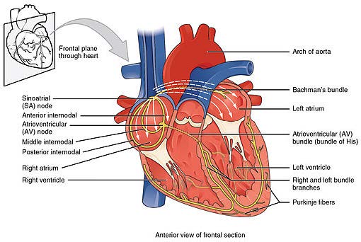
As previously stated, the heart is a mechanical pump driven by electricity. The heart has its own electrical impulses that travel along specialised pathways. The heart contracts and relaxes to pump blood around the body. The pumping action is possible because of electrical impulses that pass through the heart muscle (Marieb & Hoehn, 2010).
The SA node releases electrical stimuli at a regular rate, the rate is dictated by the needs of the body. Electrical stimulus passes through the myocardial cells of the atria creating a wave of contraction which spreads rapidly through both atria (Hatchett and Thompson, 2007). In other words, think of a wave of dominoes passing through the heart. The electrical stimulus from the SA node reaches the AV node and becomes delayed briefly. This allows the contracting atria have enough time to pump all the blood into the ventricles. Once the atria are empty, the valves between the atria and ventricles close. At this point the atria begin to refill and the electrical stimulus passes through the AV node and bundle of His into the bundle branches and Purkinje fibres (Hatchett and Thompson, 2007).
Then the ventricles contract – the right pumps blood to the lungs and the left pumps blood into the aorta. At this point the ventricles are empty, the atria full, the valves between them closed and the SA node is about to release another electrical stimulus.
Simple? But wait there is more
There is another part to this process. The SA node and AV node contain only one stimulus. Therefore every time the nodes release a stimulus they must recharge before they can do it again. The SA node recharges while the atria refill. The AV node recharges when the ventricles are refilling (Hatchett and Thompson, 2007). Hence, there is no need for a pause in heart function. All of this in less than one third of a second. Pretty impressive!
Terms Used
The term used for the release (discharge) of an electrical stimulus is depolarisation and the term for recharging is repolarisation.